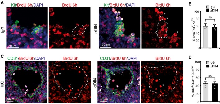
Figure EV3. Proliferation of IAHCs and endothelial cells is not changed upon αDll4 treatment.

-
A, CRepresentative confocal images of transversal sections in the AGM region. Autofluorescent circulating cells are signed by asterisks. Scale bar: 30 μm.
-
B, DBars represent the percentage of Kit+ or CD31+ cells that incorporated BrdU. Mean ± SE (n = 3).
