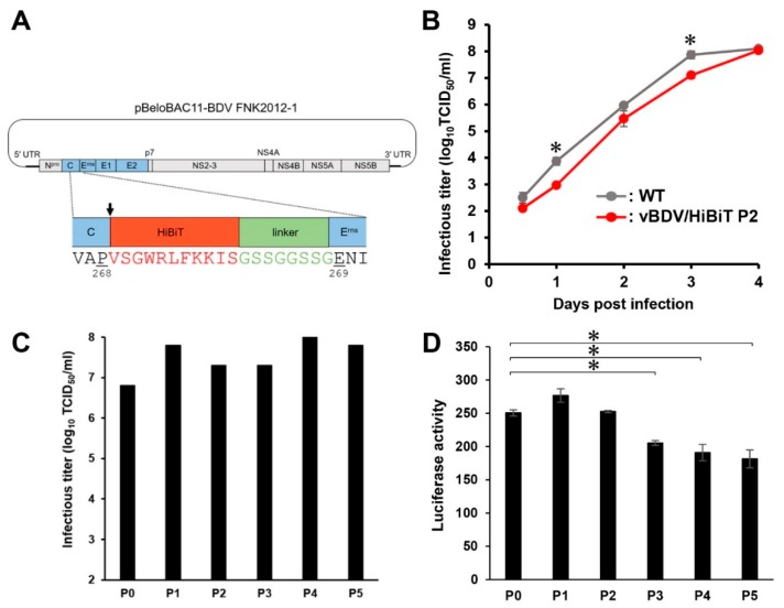
Figure 1.
Construction and characterization of recombinant border disease virus (BDV) carrying high-affinity NanoBiT (HiBiT). (A) Schematic diagram of the BDV cDNA clone encoding the HiBiT luciferase gene. Amino acid sequence of HiBiT (red) and linker (green) was inserted upstream of Erns of BDV FNK2012-1 strain (indicated by arrow). (B) Swine kidney line-L (SK-L) cells were infected with 200 50% tissue-culture infective doses (TCID50) of BDV FNK-WT (WT) and the recombinant vBDV FNK/HiBiT P2 (vBDV/HiBiT P2). Infectious titers in the culture supernatant were determined at the indicated time points. Data are presented as mean ± standard error (SE) (n = 3). Asterisks indicate significant differences (*, p < 0.05) versus the result of the vBDV/HiBiT P2. (C) SK-L cells were infected with 200 TCID50 of vBDV FNK/HiBiT, then the recombinant BDV was passaged five times. Undiluted culture supernatant (100 µL) at 3 days post-infection (dpi) was used for the next passage in naïve cells, and infectious titers in the culture supernatant were determined at 4 dpi after each passage. (D) SK-L cells were infected with 200 TCID50 of vBDV FNK/HiBiT, then the recombinant BDV was passaged five times. Luciferase activity in the cells was determined at 4 dpi after each passage. Data are presented as mean ± standard error (SE) (n = 3). Asterisks indicate significant differences (*, p < 0.05) versus the result of the vBDV FNK/HiBiT P0.

