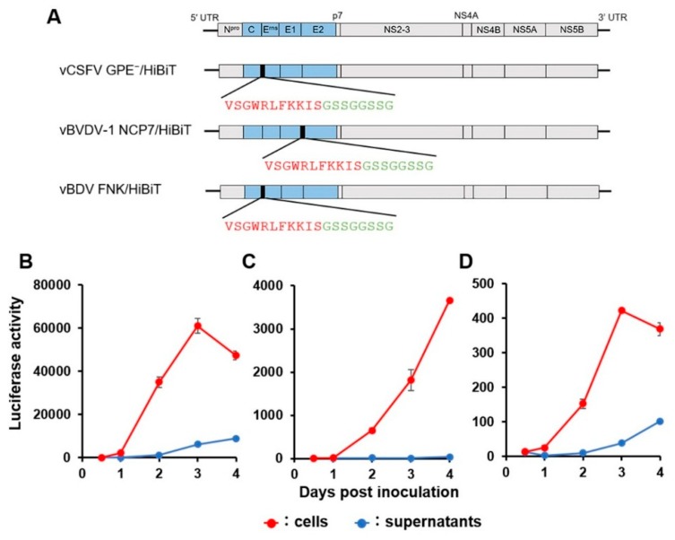
Figure 2.
Construction and luciferase activity of the recombinant pestiviruses carrying HiBiT. Schematic preparation of the reporter pestiviruses (A). Coding regions of the recombinant viruses are displayed as boxes divided by each protein gene; structural protein (blue) and non-structural protein (grey). Amino acid sequence of HiBiT (red) and linker (green) was inserted upstream of Erns of classical swine fever virus (CSFV) and BDV or upstream of E2 of BVDV-1. The luciferase activities following the infection of recombinant vCSFV GPE−/HiBiT in swine kidney line-L (SK-L) cells (B), vBVDV-1 NCP7/HiBiT in Madin–Darby bovine kidney (MDBK) cells (C), and vBDV FNK/HiBiT in SK-L cells (D) at 200 TCID50 were determined in the cell lysate or the culture supernatant, respectively. Data are presented as mean ± standard error (SE) (n = 3).

