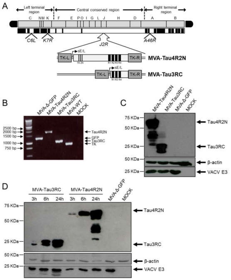
Figure 1.
Generation and in vitro characterization of MVA-Tau4R2N and MVA-Tau3RC vaccine candidates. (A) Scheme of the MVA-Tau4R2N and MVA-Tau3RC genome map. The full-length human tau gene (isoform Tau4R2N, including 2 N-terminal inserts and 4 microtubule-binding repeats) and the Tau3RC fragment (including 3 microtubule-binding repeats) are driven by the VACV sE/L promoter and are inserted within the VACV TK viral locus (J2R). The deleted VACV C6L, K7R, and A46R genes are indicated. TK-L, TK left; TK-R, TK right. (B) PCR analysis of the VACV TK locus. Viral DNA was extracted from DF-1 cells mock infected or infected at 5 PFU/cell for 24 h with MVA-Tau4R2N, MVA-Tau3RC, MVA-Δ-GFP, or MVA-WT. Primers spanning the TK locus-flanking regions were used for PCR analysis of the human Tau4R2N and Tau3RC genes inserted within the VACV TK locus. DNA products are indicated by an arrow on the right. A molecular size marker (1-kb ladder) with the corresponding sizes (base pairs) is indicated on the left. (C) Expression of human Tau4R2N and Tau3RC proteins. DF-1 cells were mock infected or infected at 5 PFU/cell with MVA-Tau4R2N, MVA-Tau3RC, or MVA-Δ-GFP. At 24 hpi, cells were lysed, fractionated by 10% SDS-PAGE, and analyzed by Western blotting. Arrows on the right indicate the positions of the full-length human Tau4R2N or the Tau3RC proteins (detected with the anti-tau antibody 7.51), VACV E3 protein or β-actin. The sizes of standards (in kDa) are indicated on the left. (D) Expression kinetics of human Tau4R2N and Tau3RC proteins. DF-1 cells were mock infected or infected at 5 PFU/cell with MVA-Tau4R2N, MVA-Tau3RC or MVA-Δ-GFP. At 3, 6 and 24 hpi expression of full-length human Tau4R2N and Tau3RC proteins, VACV E3 protein or β-actin was determined in cell extracts by western blot, as above.

