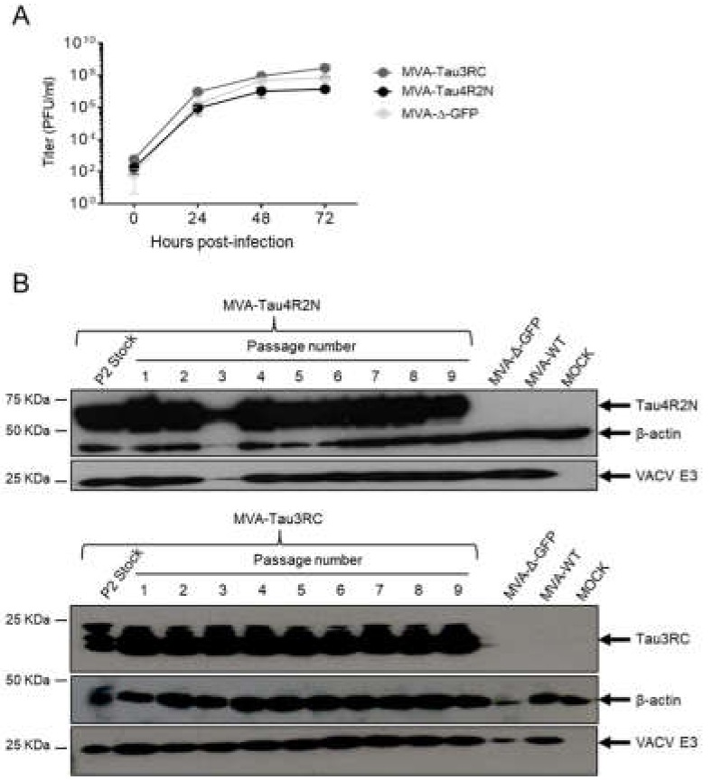
Figure 2.
Viral growth kinetics and stability of MVA-Tau4R2N and MVA-Tau3RC. (A) Viral growth kinetics of MVA-Tau4R2N and MVA-Tau3RC. Monolayers of DF-1 cells were infected at 0.01 PFU/cell with MVA-Tau4R2N, MVA-Tau3RC, or MVA-Δ-GFP. At different times postinfection (0, 24, 48, and 72 hpi), virus titers in cell lysates were quantified by a plaque immunostaining assay. The mean and standard deviations from two independent experiments are shown. (B) Stability of MVA-Tau4R2N and MVA-Tau3RC. P2 stocks of MVA-Tau4R2N and MVA-Tau3RC were continuously grown at low MOI of 0.01 PFU/cell to passage 9 in DF-1 cells. The expression of Tau4R2N and Tau3RC proteins was determined after 3 days of infection by Western blotting using the anti-tau antibody 7.51. Rabbit anti-β-actin and anti-VACV E3 protein antibodies were used as a cellular and VACV loading controls, respectively. Arrows on the right indicate the position of the Tau4R2N and Tau3RC proteins, β-actin and the VACV E3 protein. The sizes of standards (in kDa) are indicated on the left.

