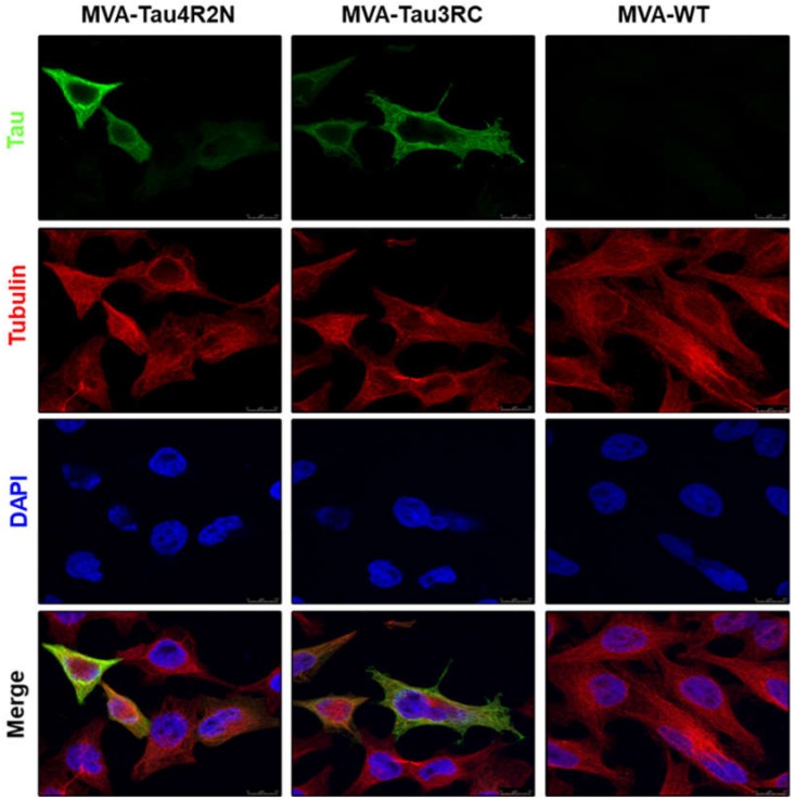
Figure 3.
Immunofluorescence analysis of human tau proteins expressed by MVA-Tau4R2N and MVA-Tau3RC. HeLa cells were infected at 0.5 PFU/cell with MVA-Tau4R2N, MVA-Tau3RC, or MVA-WT for 24 h. Then, cells were fixed and permeabilized, followed by labeling with a mouse monoclonal anti-tau antibody (7.51), or a rabbit anti-tubulin antibody. Anti-tau was detected with a mouse secondary antibody conjugated with the fluorochrome Alexa Fluor 488 (green), and anti-tubulin was detected with a rabbit secondary antibody conjugated with Alexa Fluor 594 (red). Cell nuclei were stained using DAPI (blue). Scale bar: 10 μm.

