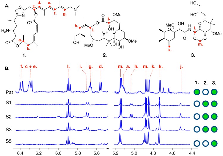
FIG 1.
Structures of major metabolites and chemotyping of Mycale hentscheli specimens. (A) Structures for each of the cytotoxic polyketides previously isolated from M. hentscheli are shown. These are pateamine A (compound 1), peloruside A (compound 2), and mycalamide A (compound 3). Red labels indicate positions of protons whose shifts were diagnostic of compound presence during chemotyping experiments. (B) Selected regions of 1H NMR spectra for each of the five specimens examined in this study. Diagnostic peaks for the presence of each compound are labeled with dashed red lines, and the letters above these match the positions of protons in panel A. The right-hand panel indicates the compounds that were determined to be present in each specimen.

