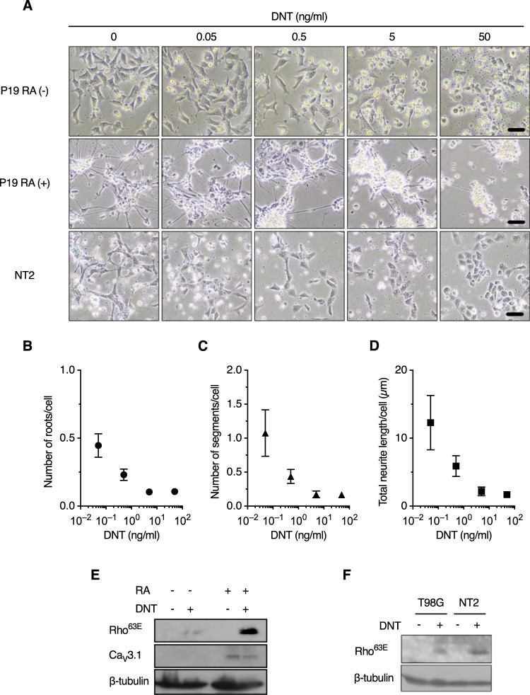
FIG 4.
Neural cells are sensitive to DNT. (A) Morphological changes of P19 {undifferentiated [RA (−)] and differentiated [RA (+)]} and NT2 cells. The cells were treated with DNT at the indicated concentrations for 24 h. Bars, 50 μm. (B to D) Numbers of nerve roots and neurite segments and total length of neurites of P19 RA (+) cells (see also Fig. S3C in the supplemental material). The cells were treated with DNT at the indicated concentrations for 24 h in a 12-well plate, and the numbers of nerve roots (B) and neurite segments (C) and the total length of neurites (D) were evaluated from 4,000 to 25,000 cells in a single well using the Opera Phenix system and Harmony 4.5. Values are means ± SEM (n = 4). (E and F) Immunoblotting of P19 cells (E) and T98G and NT2 cells (F) for deamidated Rho. The cells were treated with 50 ng/ml of DNT for 24 h.

