FIG 6.
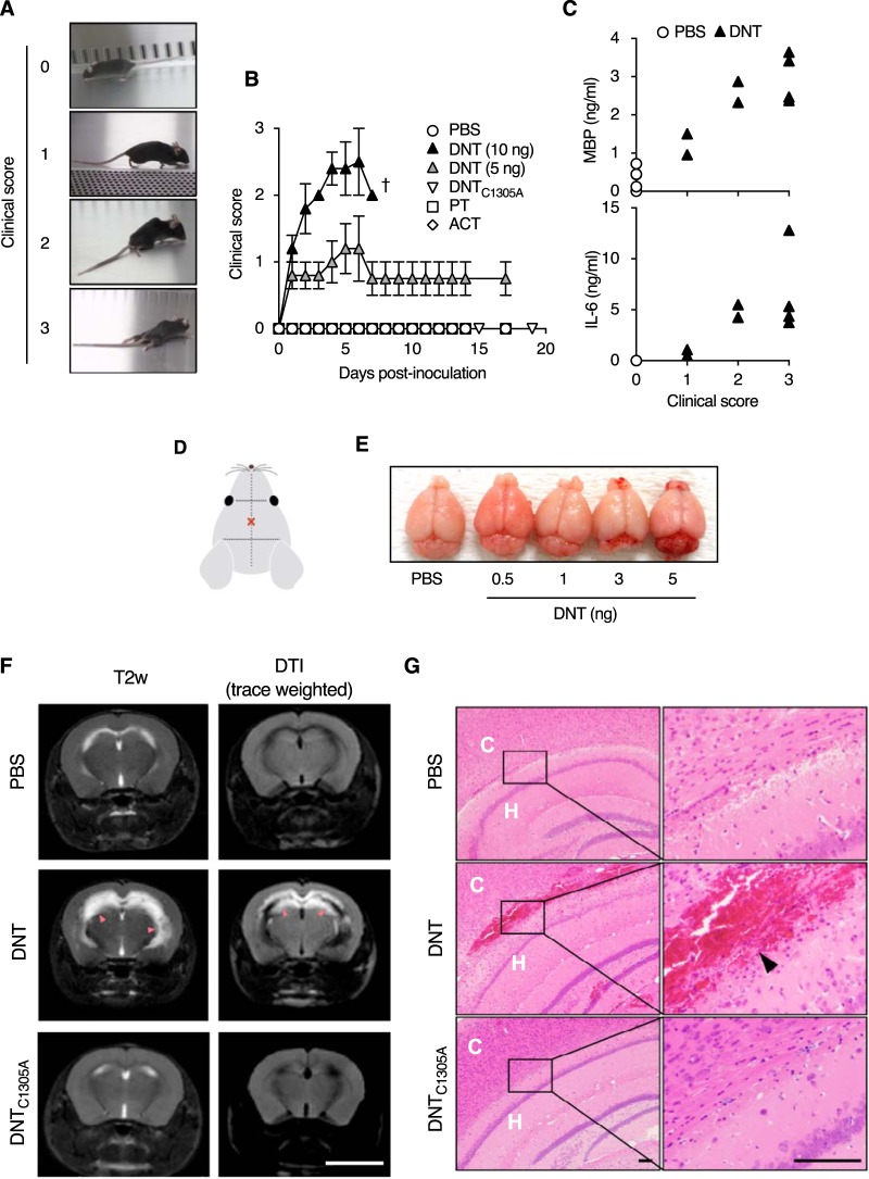
DNT-induced encephalitis in mice. (A and B) Clinical signs of neurological disorders in mice intracerebrally injected with DNT, DNTC1305A, PT, and ACT. The severities of signs were scored as follows: 0 for normal, 1 for limp tail and hind limb weakness, 2 for partial paralysis of hind limbs, and 3 for complete paralysis of hind limbs. Each plot represents means ± SEM (n = 5). †, all dead. One and two mice died 5 days and 13 days after injection with PT, respectively. (C) Concentrations of MBP (top) and IL-6 (bottom) in cerebrospinal fluids of mice intracerebrally injected with DNT (n = 8) or PBS (n = 5). Each data point, representing one mouse, was plotted on the ordinate against each clinical score on the abscissa. (D) Illustration indicating the injection site (×). Samples were injected with a needle 4 mm long and 0.4 mm in diameter. (E) Macroscopic images of mouse brains excised 3 days after DNT injection. Note the bloody spots around the cerebellum and olfactory bulb. (F) MRI images of brains of mice intracerebrally injected with 5 ng of DNT or DNTC1305A. The mice were subjected to MRI 3 days after injection. Bar, 5 mm. T2-weighted (T2w) imaging and diffusion tensor imaging (DTI) revealed abnormal high-intensity white signals (arrowheads) in the vicinity of cerebral ventricles of the DNT-injected mice, indicating severe inflammation (DTI) with watery infiltration (T2w). (G) Histological sections of brains of mice intracerebrally injected with 3 ng of DNT or 5 ng of DNTC1305A. Hemorrhage is noted in the specimen of a mouse injected with DNT (arrowhead). Bars, 100 μm. C, cerebral cortex; H, hippocampus.

