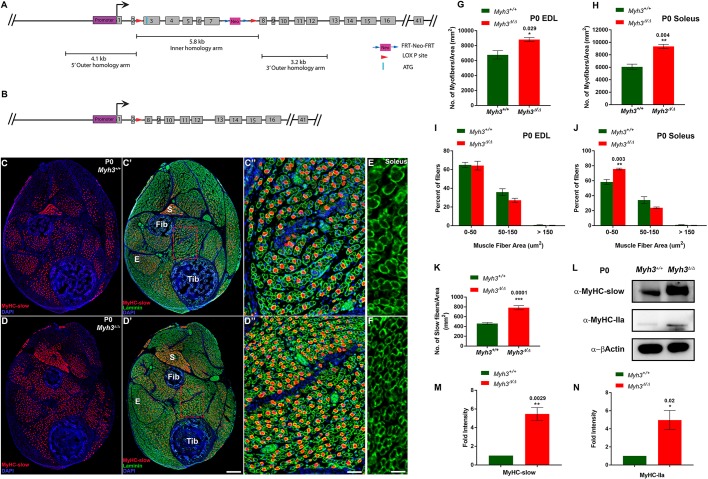
Fig. 1.
Loss of MyHC-embryonic leads to neonatal myogenic differentiation defects. (A,B) Schematics depicting the Myh3fl3-7 allele, where LoxP sites (red arrowheads) flank exons 3-7 (A) and the Myh3Δ allele lacking exons 3-7 of Myh3 (B). (C-D″) Cross-sections through the hind limbs of P0 Myh3+/+ (C,C′) and Myh3Δ/Δ (D,D′) mice labeled by immunofluorescence for MyHC-slow (red), laminin (green) and DAPI (blue). ‘E’ and ‘S’ denote EDL and soleus muscles, respectively, and ‘Tib’ and ‘Fib’ denote tibia and fibula bones, respectively, in C′ and D′; C″ and D″ are magnifications of boxed areas of the flexor digitorum longus (FDL) muscle from C′ and D′, respectively. (E,F) Laminin (green) labeling on sections through the soleus muscles of P0 Myh3+/+ (E) and Myh3Δ/Δ (F) mice. (G,H) Quantification of myofiber number through the EDL (G) and soleus (H) muscles of P0 Myh3+/+ and Myh3Δ/Δ mice. (I,J) Quantification of myofiber area through the EDL (I) and soleus (J) muscles of P0 Myh3+/+ and Myh3Δ/Δ mice, grouped into 0-50 µm2, 50-150 µm2 and above 150 µm2. (K) Quantification of MyHC-slow+ fibers from P0 Myh3+/+ and Myh3Δ/Δ shank whole cross-section normalized to total area. (L-N) Western blots for MyHC-slow, MyHC-IIa and β-actin from P0 Myh3+/+ and Myh3Δ/Δ mice hind limb protein lysates (L) and densitometric quantification (M,N). Data are mean±s.e.m. of a minimum of three independent experiments. Scale bars: 200 μm (D′); 40 μm (D″); 20 μm (F).

