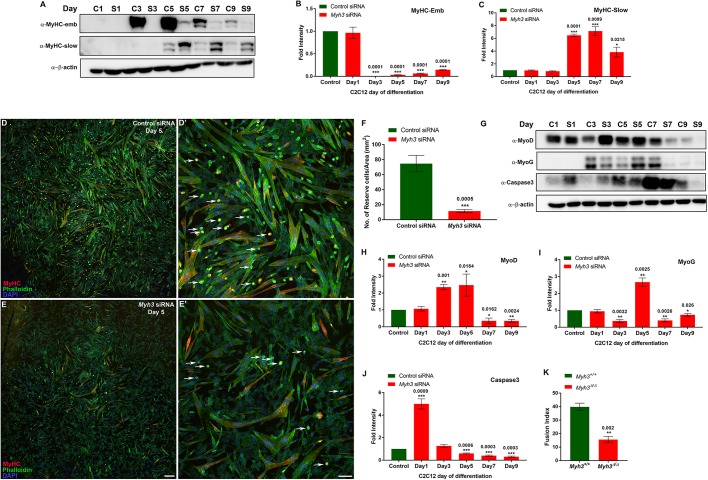
Fig. 5.
Myh3 depletion causes reduction in reserve cell number and decreased fusion index. (A-C) Western blots for MyHC-emb, MyHC-slow and β-actin on control and Myh3 siRNA-treated C2C12 cells over 9 days of differentiation (A), and densitometric quantification (B,C); ‘C’ and ‘S’ denote control and Myh3 siRNA along with the specific day of differentiation (A). (D-E′) MyHC (red), phalloidin (green) and DAPI (blue) immunofluorescence on control (D,D′) and Myh3 (E,E′) siRNA-treated C2C12 cells at day 5 of differentiation (D′ and E′ are magnifications from D and E, respectively, with white arrows marking reserve cells). (F) Quantification of reserve cell number per unit area (mm2) from control and Myh3 siRNA-treated C2C12 cells. (G-J) Western blots for MyoD, myogenin, caspase 3 and β-actin on control and Myh3 siRNA-treated C2C12 cells during differentiation (G) and densitometric quantification (H-J); ‘C’ and ‘S’ denote control and Myh3 siRNA along with the specific day of differentiation (G). (K) Fusion index of myofibers formed by differentiating myoblasts for 7 days, isolated from Myh3+/+ and Myh3Δ/Δ P0 mice, for which representative images are shown in Fig. S5C,D. Data are mean±s.e.m. of a minimum of three independent experiments. Scale bars: 100 μm (E); 25 μm (E′).

