Fig. 1.
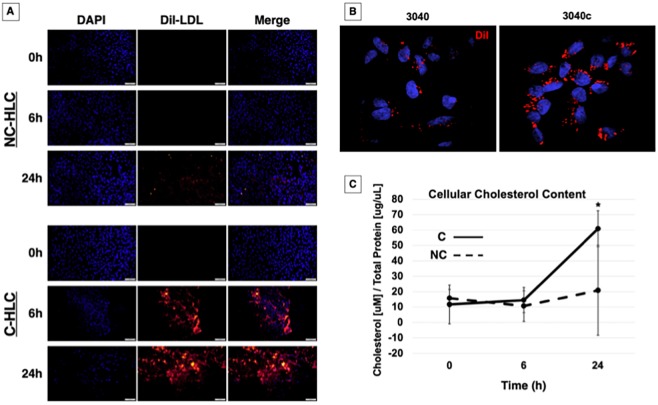
LDLR-mediated cholesterol internalization is restored in C-HLC. (A) NC-HLC and C-HLC were treated overnight in 5% LPDS medium supplemented with rosuvastatin. This was followed by incubation with DiI-LDL for 6 h or 24 h. NC-HLC did not show any DiI fluorescence from LDL uptake until 24 h. C-HLC internalized DiI-LDL by 6 h, which increased after 24 h. Scale bars: 100 μm (each image has been linearly brightened to the same level). (B) HLC were imaged at 100× magnification for detection of DiI fluorescence localization. (C) HLC were treated with LPDS medium and rosuvastatin for 48 h followed by incubation in methyl-β-cyclodextrin (MBC) for 45 min when time point 0 h samples were collected. Cells were further incubated for 6 h or 24 h with unlabeled LDL then analyzed for cellular cholesterol concentration with respect to protein. NC-HLC showed no statistical difference in cellular cholesterol for all three time points, demonstrating the dysregulation of cholesterol metabolism in FH HLC. Corrected cells did not have statistically different cholesterol content after 6 h, demonstrating the effect of MBC treatment and plasma membrane cholesterol chelation on receptor-medicated endocytosis. After 24 h exposure to LDL-C, C-HLC were able to increase cellular cholesterol to a statistically significant level [61 µM/(µg/µl) compared to 11.6 µM/(µg/µl) at 0 h]. There was no difference in cholesterol concentration at 0 h between NC-HLC and C-HLC. The graph values represent the mean±s.d. (n=3) per treatment, per cell type and replicated three times in the laboratory. Statistics were performed using a two-way ANOVA Holm–Sidak post-hoc test. *P<0.05 compared to NC-HLC at 6 h and 24 h, and C-HLC at 6 h.

