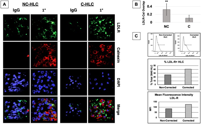
Fig. 2.
LDLR colocalizes with calnexin-ER in FH HLC. (A) After differentiation, HLC were treated overnight in LPDS medium supplemented with rosuvastatin followed by immunocytochemistry and confocal imaging, 100× oil immersion objective. AMIRA software was used to stack slices and merge channels to present an overview of localization of the LDLR (green), calnexin-ER (red) and nucleus (DAPI). (B) Quantification of colocalization using Fluoview software indicated that NC-HLC did have a significantly greater colocalization of the LDLR with calnexin than C-HLC. The graph values represent the mean±s.d. (n=5) per cell type from three experiments repeated in the laboratory. Statistics were performed using an unpaired two-tailed Student's t-test, **P<0.01. (C) Both non-corrected (NC) and corrected (C) cell lines were differentiated to HLC and assayed for surface LDLR expression using AF647-conjugated anti-LDLR antibody clone C7 (top). Cells were quantified for percentage total positive labeling (middle) and mean fluorescence intensity (MFI) (bottom).

