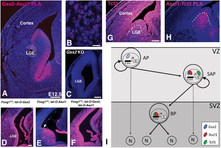
Fig. 8.
Proximity ligation assay (PLA) shows Ascl1-Gsx2 interactions and Ascl1-Tcf3 interactions in a distinct portion of the LGE germinal zone. (A) When PLA was performed using rabbit anti-Gsx2 and guinea pig anti-Ascl1 antibodies, a strong signal (magenta) was detected in the LGE and septal VZ. (B) High power magnification of the VZ region shows a punctate signal associated with the DAPI-stained nuclei. (C) The PLA signal between Gsx2 and Ascl1 antibodies was specific as no signal was detected in Gsx2 knockout (KO) tissue sections. (D) In Gsx2-misexpressing embryos, PLA signal is expanded throughout the telencephalon as is the case for Gsx2 and Ascl1 expression. (E) PLA signal is not expanded throughout the telencephalon in the Ascl1-misexpressing embryos (pallio-subpallial boundary indicated by arrow in E as Gsx2 is not upregulated outside of the ventral telencephalon (Fig. 2B). However, the PLA signal is intensified in the ventral telencephalon. (F) Misexpression of both Gsx2 and Ascl1 leads to increased PLA signal throughout the telencephalon. (G) Immunostaining for Tcf3 protein in the E12.5 telencephalon shows staining throughout the germinal zones including both the VZ and SVZ. (H) PLA using the goat anti-Tcf3 and guinea pig anti-Ascl1 antibodies shows signal in both the LGE VZ as well as the SVZ, with stronger signal in the latter region. The boundary between the VZ and SVZ is indicated by the dashed line in G and H. (I) Schematic model showing LGE progenitor subtypes, with SAPs co-expressing Gsx2 and Ascl1 thus limiting Ascl1's neurogenic function and allowing for progenitor expansion. Gsx2 expression is lost in BPs, allowing Ascl1:Tcf3 heterodimers to drive direct neurogenesis. Note that Gsx2 was observed in some APs (indicated by blue hatching) but not together with Ascl1. In this model, both APs and SAPs could undergo direct neurogenesis if Gsx2 was downregulated (indicated by thin arrows). Scale bars:100 µm (A); 10 µm (B); 200 µm (C); 100 µm (D-H). N, neuron.

