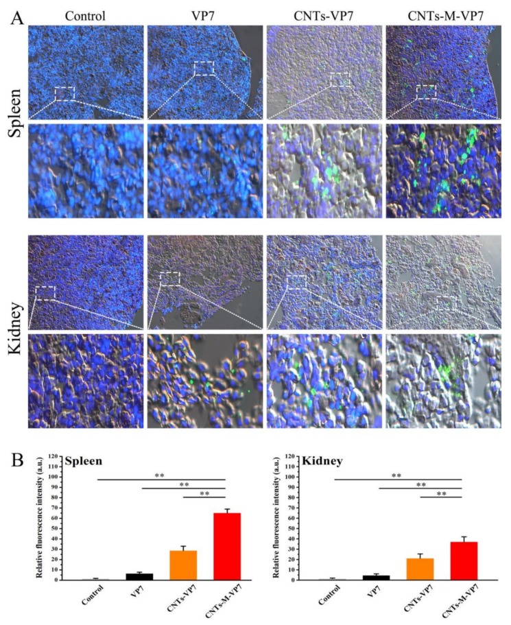
Figure 3.
Uptake of nanovaccine in fish tissues. (A) The immunofluorescence images of fish tissues (spleen and kidney) after incubated with vaccines (VP7, CNTs-VP7, and CNTs-M-VP7), respectively. The vaccines were labeled with FITC (green channel); The cell nucleus was labeled with DAPI (blue channel). (B) Mean fluorescence intensity of vaccine in fish treated with VP7 (black column), CNTs-VP7 (Orange column), CNTs-M-VP7 (Red column). Free FITC treatment was conducted as the control (light grey column). Data are presented as the mean ± SD. p values were calculated by Student’s t-test (** p < 0.01, * p < 0.05).

