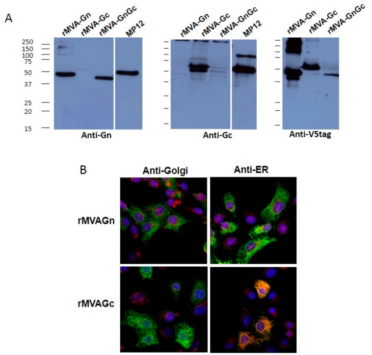
Figure 1.
Expression and subcellular localization of recombinant Gn and Gc upon modified vaccinia Ankara (MVA) infection. (A). Western blot of different MVA infected BHK-21 cell extracts probed with mAb 84a antiGn or a rabbit polyclonal serum antiGc. The antiV5 tag mAb was used to compare the Gn and Gc expression levels and to confirm the expression of the full-length antigen. As a positive control a RVFV-MP12 infected cell extract was used. Numbers indicate relative molecular mass in kilodaltons. (B). Confocal immunofluorescence images of MVA infected Vero cells. Expression of Gn or Gc was detected with anti V5 tag mAb (green). Intracellular membranes were labeled with either antihuman mannosidase-II (Golgi) or anticalreticulin (ER) mAbs (red fluorescence) as indicated. Nuclei were labeled with DAPI stain (blue). All panels correspond to merged fluorescence images. Colocalization of Gc and ER membranes is evidenced by yellow-orange fluorescence.

