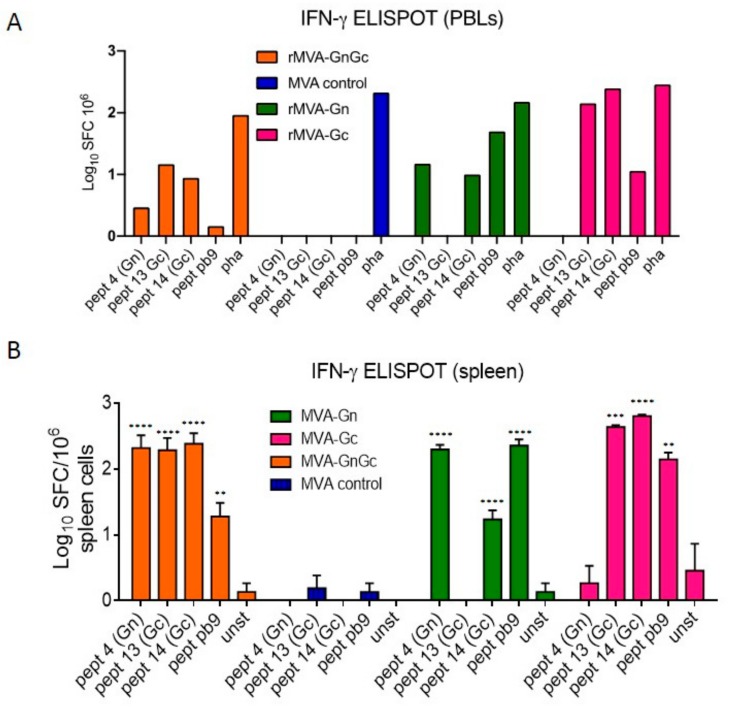
Figure 4.
Cellular responses upon MVA immunization. (A). Interferon gamma ELISPOT assay of pooled (n = 5) BALB/c peripheral blood leukocytes (PBLs) taken at day 14 postimmunization with the different MVA vaccines. Each pool was restimulated with either Gn (#4), or Gc (#13 or #14) specific peptides or with peptide pb9. Nonspecific stimulation was induced with phytohemaglutinin (pha). (B). Mean ± SD log spot forming cells (SFC) values obtained in spleen cells from BALB/c mice (n = 2) at day 7 post MVA immunization. As above, the peptides 4, 13, and 14 were selected on the basis of their ability to stimulate Gn and Gc specific T-cell responses. Cell culture medium with no added peptide (unst) was used to measure the background of the assay. The pb9 peptide was used as a specific positive control for each recombinant MVA (rMVA) vaccinated mice. In all groups asterisks indicate significance levels for each peptide when compared to the unstimulated control (unst) using Dunnett’s multiple comparisons test (** p < 0.01; *** p < 0.001; **** p < 0.0001).

