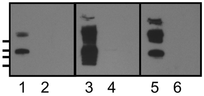
Figure 2.
Western blot analysis of spore surface extracts. Lanes 1, 3, and 5—Clostridium difficile R20291 spore surface extract; lanes 2, 4, and 6—R20291::sgtA spore surface extract. Lanes 1 and 2 reacted with anti-β-O-linked GlcNAc antibody, lanes 3 and 4 reacted with rabbit polyclonal anti KLH-SML10 glycopeptide antiserum, lanes 5 and 6 reacted with rabbit anti-formalin killed spore antiserum (CD5). Molecular weight markers indicated on left hand side (top to bottom) 250, 150, 100, and 75 kDa.

