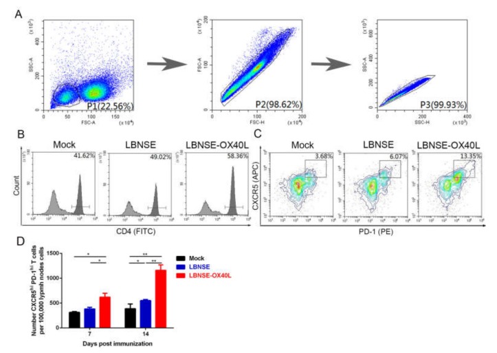
Figure 3.
Generation of T follicular helper (Tfh) cells in mice immunized with LBNSE-OX40L. Moreover, 105 single inguinal lymph node (LN) cells from the mice immunized with 106 FFU of LBNSE, LBNSE-OX40L, or DMEM (as mock) were stained by antibodies for analyzing the Tfh cells (CD4+CXCR5hiPD-1hi) by flow cytometry. Cell populations of interest were visualized by pseudocolor plots (A) or histogram plots (B) or contour plots (C) showing their forward-scatter (FSC) and side-scatter (SSC) signals in relation to the size and granularity/complexity of the cells, respectively. The gating strategies and representative contour plots of lymphocyte populations in LN cells (A), CD4+T cell populations in lymphocyte (B), and Tfh cell populations in CD4+T cells (C) were shown. (D) At 7 and 14 dpi, the total number of Tfh cells per 105 draining LN cells was determined. Error bars represented the SE (n = 3). The following notations were used to indicate significant differences between groups: *, p < 0.05; **, p < 0.01.

