Figure 4.
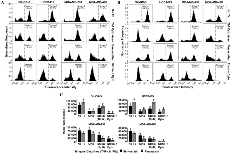
Statin drugs plus Th1 cytokines maximize mitochondrial membrane depolarization as assessed by tetramethylrhodamine ethyl ester (TMRE) staining. SK-BR-3, HCC1419, MDA-MB-231, and MDA-MB-468 human breast cancer cell lines were cultured with no additives (No Tx), treated with recombinant Th1 cytokines (TNF-α and IFN-γ, 10 ng/mL each), a statin drug (A) Simvastatin or (B) Fluvastatin (1 µM MDA-MB-231; 10 µM remaining cell lines), or the combination of Th1 cytokines and a statin drug (A) “Simva + Cyto” or (B) “Fluva + Cyto” for approximately 48 h. Flow cytometric results displayed in panels A and B are from one representative experiment. (C) Statistical analysis of flow cytometry results comparing the mean channel fluorescent intensity between groups: no additives (No Tx), treated with recombinant Th1 cytokines (TNF-α and IFN-γ, 10 ng/mL each), a statin drug (Simvastatin or Fluvastatin, 1 µM MDA-MB-231; 10 µM remaining cell lines), or the combination of Th1 cytokines and a statin drug (“Statin + Cyto”). Results displayed are from at least five trials +/− SEM. Letter designations represent Tukey’s HSD comparisons: treatments with the same letter designation are not statistically different; when letter designations differ between treatments, the p-value is less than 0.05.

