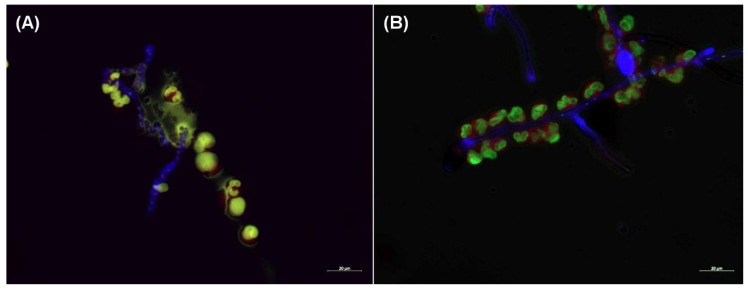
Figure 3.
Neutrophil extracellular trap (NET) formation is missing when neutrophils are confronted with the hyphae of C. lunata. Fluorescent microscopy was performed about after 3 h of interaction of serum opsonized A. fumigatus (A) and C. lunata (B) with neutrophil granulocytes. Hyphae and conidia were labeled with Calcofluor white (blue); neutrophil’s cell membrane was stained with CellMask Deep Red Plasma Membrane Stain (red) and the DNA was stained with SYTOX Green (green). NET formation was detected in the interaction with A. fumigatus but only gathering of the cells to the hyphae could be observed in case of C. lunata.

