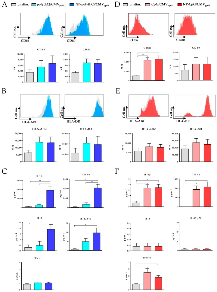
Figure 3.
Maturation of conventional DCs (cDCs) and plasmacytoid DCs (pDCs) by functionalized CaP nanoparticles. (A) Representative histograms show expression of costimulatory molecules cDCs. Blood mononuclear cells (PBMC) isolated cDCs were stimulated with poly(I:C) functionalized CaP NPs or soluble poly(I:C). After 24 h expression of costimulatory molecules, CD86 and CD80 was analyzed by flow cytometry. The mean fluorescence intensity (MFI) is depicted. (B) 24 h after stimulation expression of MHC I (HLA-ABC) and MHC II (HLA-DR) was analyzed by flow cytometry. (C) Supernatants from stimulated cDCs were analyzed for proinflammatory cytokines after 24 h. (D) PBMC-isolated pDCs were stimulated with CpG functionalized CaP NPs or soluble CpG. After 24 h expression of costimulatory molecules, CD86 and CD80 was analyzed by flow cytometry. (E) MHC I (HLA-ABC) and MHC II (HLA-DR) expression was analyzed by flow cytometry. (F) Supernatants from stimulated pDCs were investigated for proinflammatory cytokines after 24 h of incubation. Results are summarized from at least three independent experiments. Bars represent means with SEM. One-way ANOVA was used, which was followed by the Tukey’s multiple comparisons test. * p < 0.05, ** p < 0.01.

