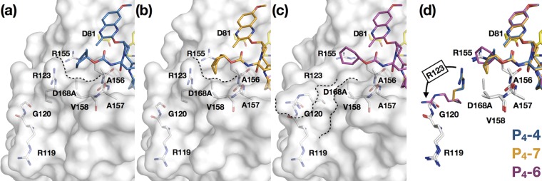
FIG 6.
Fit of P4 capping groups and the conformation of R123 reshaping the S4 pocket of HCV NS3/4A protease. P4-4 (a), P4-7 (b), and P4-6 (c) cocrystal structures are shown with the D168A variant. The protease is in surface representation, with residues in and around the S4 pocket (white) and the catalytic triad (yellow) side chains in stick representation. The contour of the S4 pocket is outlined in dotted lines. (d) Superposition of P4-4, P4-7, and P4-6, as indicated, showing the alternate conformations of Arg123 (in respective color of the inhibitors) in the cocrystal structures.

