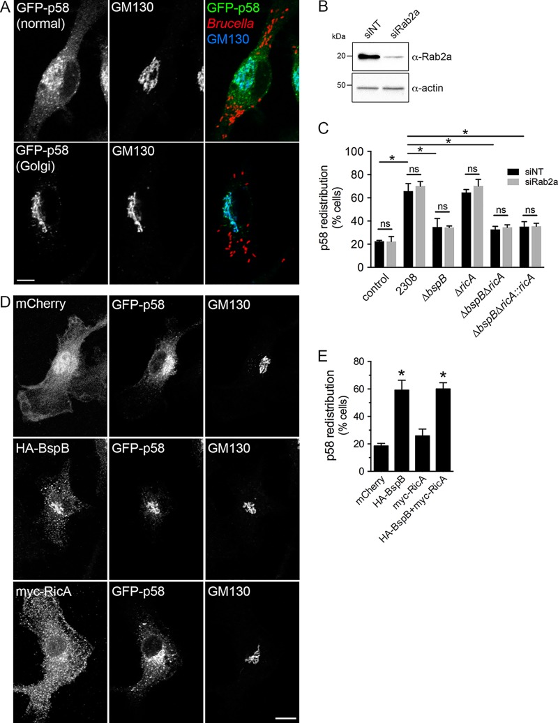
FIG 4.
BspB-mediated alterations of ER-to-Golgi transport are independent of RicA and Rab2a. (A) Representative confocal micrographs of GFP-p58 steady-state distribution in Brucella-infected BMMs transduced to express GFP-p58, showing either normal or Golgi apparatus-localized patterns. Golgi structures were labeled using an anti-GM130 antibody. Scale bar, 10 μm. (B) Representative Western blotting of Rab2a depletion in BMMs. BMMs were nucleofected with either nontargeting (siNT) or siRab2a siRNAs, and Rab2 levels were evaluated after 72 h via Western blotting of Rab2a and β-actin as a control. (C) Quantification of GFP-p58 redistribution to the Golgi apparatus in BMMs treated with either nontargeting (siNT) or siRab2a siRNAs and then left uninfected (control) or infected for 24 h with wild-type (2308), ΔbspB, ΔricA, ΔbspB ΔricA, or ΔbspB ΔricA::ricA B. abortus strains. Values are means ± SD of results from 3 independent experiments. Asterisks indicate a statistically significant difference between tested conditions, assessed using one-way analysis of variance (ANOVA) followed by a Dunnett’s multicomparison test. ns, not significant. (D) Representative confocal micrographs of BMMs cotransduced to express GFP-p58 with mCherry, HA-BspB, or myc-RicA, showing normal or Golgi apparatus-localized distribution of GFP-p58. Scale bar, 10 μm. (E) Quantification of GFP-p58 redistribution to the Golgi apparatus in BMMs cotransduced to express GFP-p58 with mCherry, HA-BspB, myc-RicA, or HA-BspB and myc-RicA. Values are means ± SD of results from 3 independent experiments. Asterisks indicate a statistically significant difference compared to control (mCherry) cells, assessed using one-way analysis of variance (ANOVA) followed by a Dunnett’s multicomparison test. ns, not significant.

