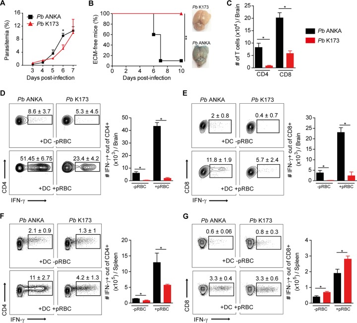
FIG 1.
Reduced Th1 responses and absence of ECM pathology following P. berghei K173 infection. C57BL/6 mice infected i.v. by injection of 106 P. berghei ANKA (Pb ANKA) or P. berghei K173 (Pb K173) pRBC. Blood circulating parasitemia (A) and ECM development (B) monitored following infection. Brain edema was visualized by Evans blue coloration. (C) Total numbers of CD4+ or CD8+ T cells collected from brain at day 6 after infection. Cells collected from brain (D and E) and spleen (F and G) at day 6 after infection were restimulated in vitro with MutuDC preloaded or not with pRBC. IFN-γ production by CD4+ T cells (D and F) or CD8+ T cells (E and G) detected by intracellular staining. Percentages on the representative dot plots show the median percentages of IFN-γ+ cells of total CD4+ or CD8+ T cells ± IQRs. Bar graphs show the medians ± IQRs of absolute numbers. Data are representative of 2 independent experiments. N = 5 mice per group.

