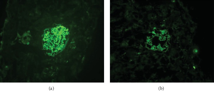
Figure 1.

Detection of podocalyxin in renal tissue using immunofluorescence methods. (a) Intense expression of podocalyxin (granular or linear pattern) in normal glomerular capillary loops. (b) In damaged glomeruli, podocalyxin immunofluorescence was reduced in intensity and uneven in distribution, with regions of deficient expression.
