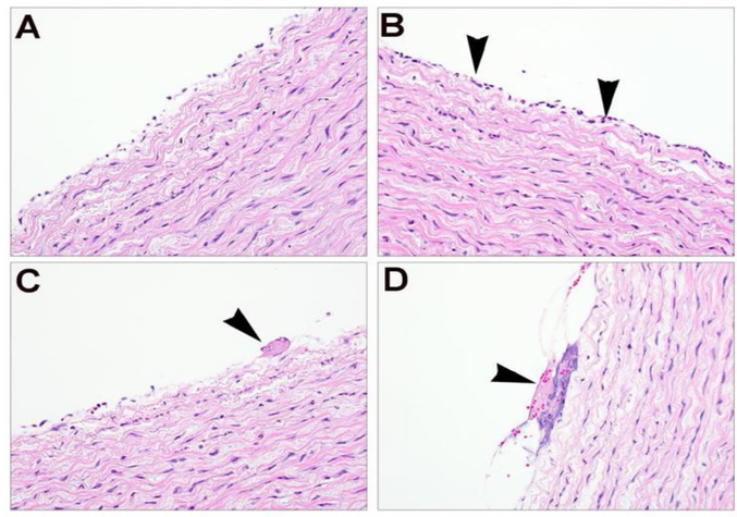
Fig. 5 -.
Representative aortic histopathology from control region (A) and treatment area (B), showing discontinuous endothelial layer in both sections, but with few scattered neutrophils (B, arrowhead) in the treated area. (C-D) Treatment areas showing few neutrophils associated with minimal accumulation of fibrin (C, arrowhead) and cellular debris (D, arrowhead).

