
Figure 3.
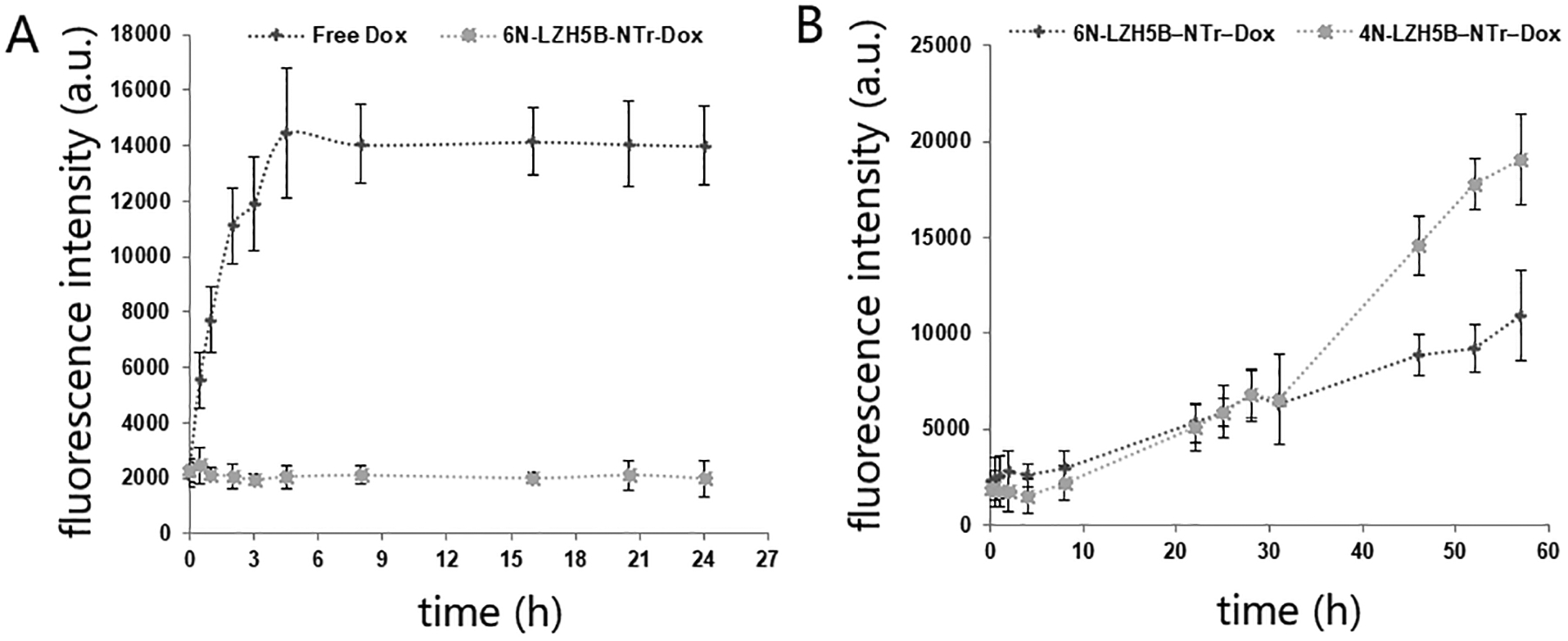
Loading and releasing of doxorubicin. Fluorescence of dialyzed doxorubicin was detected to represent the concentration of diffused doxorubicin. (A) Drug diffusion of free doxorubicin and doxorubicin carried by 6-nucleotide nanotrain in PBS with 5 mM Mg2+. (B) Drug diffusion of doxorubicin carried by 6-nucleotide nanotrain and 4-nucleotide nanotrain in PBS with 10% fetal bovine serum.