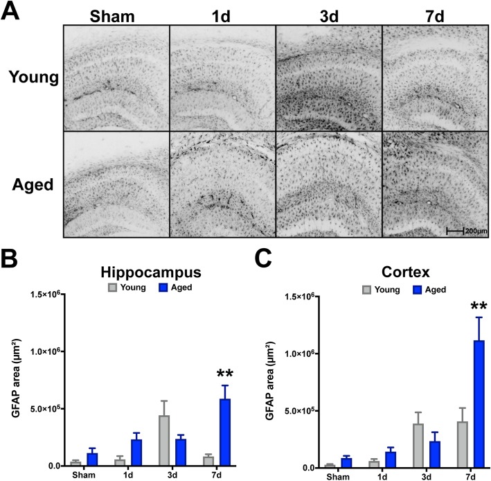
Fig. 1.
Age-related progressive reactivity of GFAP+ astrocytes in the hippocampus and neocortex after TBI. Young (4 months) and aged (18 months) mice were subjected to sham or CCI injury and euthanized at 1, 3, or 7 days post-injury. Serial sections comprising the dorsal hippocampus were imaged for GFAP labeling and quantified as the pixel positive area for each of the 8 groups. GFAP+ threshold was set on young sham tissue. TBI induced a progressive increase of GFAP+ astrocyte staining in aged mice (n = 5/group) through 7 days post-injury, whereas the GFAP+ staining intensity in young mice (n = 4/group) was most prominent by 3 days post-injury. Data were analyzed using two-way ANOVA with Sidak’s post hoc correction examining pairwise interactions for each time interval. ANOVA revealed significant differences due to age (F (1, 28) = 7.924, P = 0.0088), interval (F (3, 28) = 7.593, P = 0.0007), as well as their interaction (F (3, 28) = 9.065, P = 0.0002), for hippocampus. Similarly for the neocortex, ANOVA revealed significant differences due to age (F (1, 28) = 6.007, P = 0.0208), interval (F (3, 28) = 20.58, P < 0.0001), as well as their interaction (F (3, 28) = 6.859, P=0.0013). **P < 0.01, for pairwise comparison of 7-day interval between young and aged. Data are presented as mean ± SEM. Young, gray bars; Aged, blue bars. Scale bar is 200 μm

