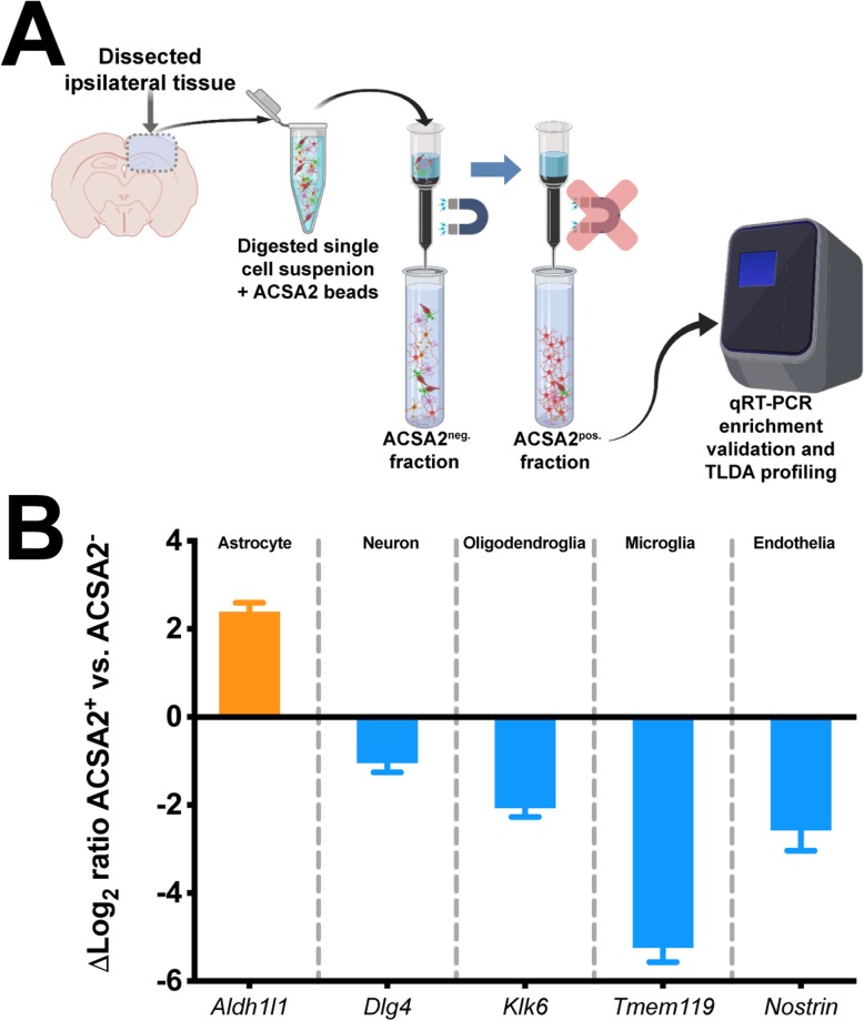
Fig. 5.
Validation of ACSA-2 astrocyte cell enrichment from brain tissue. a Generalized workflow for ACSA-2 magnetic bead enrichment of astrocytes from the injured brain parenchyma. Digested cell suspensions were labeled with the ACSA-2 magnetic bead before being placed in the magnetic column for removal of non-specific cells, which were collected into a tube as the ACSA-2neg. fraction. Removal of the column from the magnetic stand allowed the flow-through of the retained ACSA-2 astrocytes to be collected as the ACSA-2pos. fraction. RNA from both fractions was harvested to examine gene expression endpoints. b Gene expression analyses using putative markers of five neural tissue subsets: astrocyte (Aldh1l1), neuronal (Dlg4), oligodendroglial (Klk6), microglial (Tmem119), and endothelial (Nostrin). These data demonstrate a significant induction of putative astrocyte signature (orange bar), with little signature of other cell populations (blue bars). Log2 fold change is a ratio of ACSA-2pos to ACSA-2neg. TLDA, Taqman low density array cards

