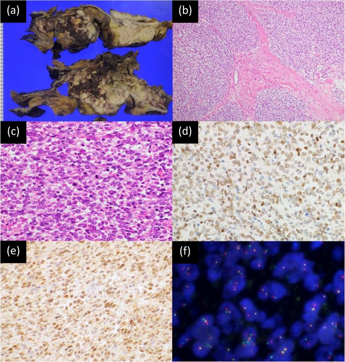
Fig. 3.
Pathological findings of the CIC-rearranged sarcoma. a Cut surface of the pancreaticoduodenectomy specimen shows a pale tan-colored tumor in the duodenum accompanied by hemorrhage and necrosis. b A low-power photomicrograph shows focal lobulation within a fibrotic stroma (20x). c A high-power photomicrograph reveals proliferation of round neoplastic cells with mild pleomorphism and focally prominent nucleoli (hematoxylin and eosin stain, 40x). d Neoplastic cells are positive for WT1 (immunohistochemistry, 40x). e Neoplastic cells are positive for ETV4 (immunohistochemistry, 40x). f Fluorescence in situ hybridization using a CIC break-apart probe set demonstrates CIC rearrangement, showing 5′ green and 3′ red abnormal split signals (100x)

