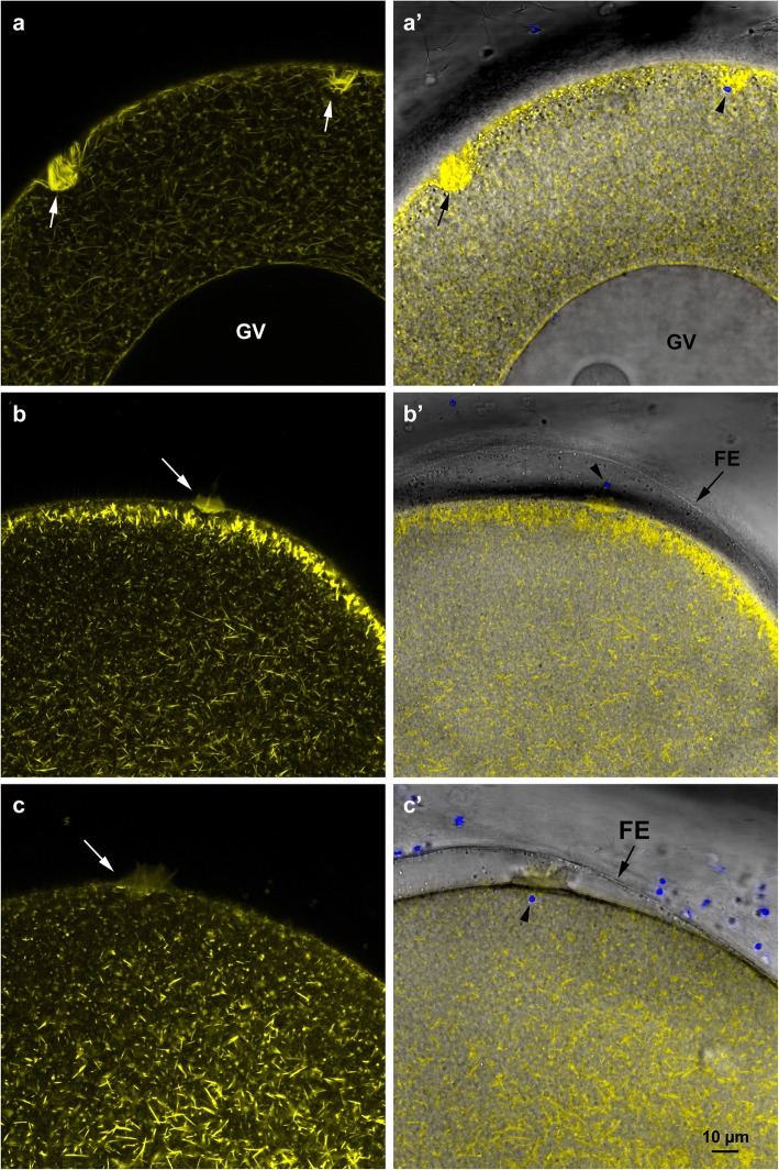
Fig. 6.
Confocal images of the cortical actin cytoskeleton in starfish oocytes fertilized at different meiotic stages. Immature oocytes, mature eggs and overripe eggs of A. aranciacus were microinjected with Alexa Fluor 568 phalloidin. a F-actin staining of the fertilization cones in a polyspermic immature oocyte. Note the large number of actin bundles composing the fertilization cones that will incorporate the sperm in the absence of a FE elevation shown in the overlay image in a’. b A mature monospermic egg fertilized in its optimal stage of maturation (70 min 1-MA treatment) that shows a fertilization cone. Note the initiation of the centripetal translocation of individual actin fibres at the subplasmalemmal zone and the elevation of the FE in the overlay image in b′ as a result of the CG exocytosis. c Reorganization of the F-actin in an overripe egg fertilized after 6 h hormonal treatment. Note that the centripetal translocation seen in fertilized mature eggs is absent in overripe eggs upon insemination. c′) Image overlay showing the fertilization envelope (FE) elevation and incorporation of sperm in the egg (arrowhead). Multiple spermatozoa enter even in the presence of normal elevation of FE

