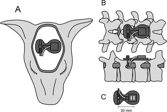
Fig. 1.
The positions of the probes, and the NIRS probe of the NIRO-200 system. A wide portion of the parietal scalp was removed, and one NIRS probe was placed on the center of the parietal skull (a). The other two NIRS probes were placed on the thoracic (not shown) and lumbar (b) laminae of the vertebral arch, which were surgically exposed and flattened after removing the spinous process. The probe consists of the emitter and receiver, housed in two aligned photodetectors, which were fixed in a rubber holder to ensure an emitter-receiver distance of 30 mm (c). Because the emitter-receiver distance was 30 mm, each spinal cord NIRS probe was placed on laminae approximately 20 mm from the center of the spinal cord

