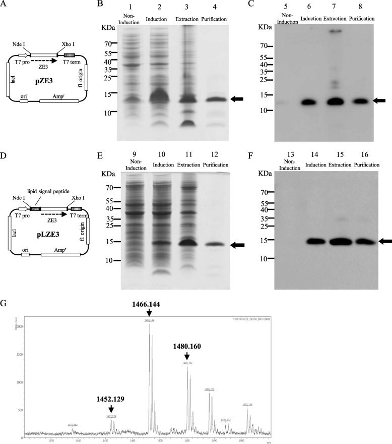
Fig. 1.
Production and purification of recombinant Zika virus envelope protein domain III (rZE3) and recombinant lipidated Zika virus envelope protein domain III (rLZE3). The plasmid maps of pZE3 (a) and pLZE3 (d) for the production of rZE3 and rLZE3, respectively. The purification of rZE3 (b, c) and rLZE3 (e, f) was monitored by 10% reducing Tricine-SDS-PAGE followed by Coomassie Blue staining and immunoblotting with anti-His-tag antibodies. rZE3 and rLZE3 were expressed in the E. coli strains BL21 (DE3) and C43 (DE3), respectively. Lanes 1, 5, 9, and 13: protein expression without IPTG induction; lanes 2, 6, 10, and 14: protein expression after IPTG induction; lanes 3 and 7: extraction of rZE3 from inclusion bodies; lanes 11 and 15: soluble fraction of rLZE3; and lanes 4, 8, 12, and 16: purified proteins. Lanes 5–8 and lanes 13–16 show the induction and purification processes for rZE3 and rLZE3, respectively, evaluated by immunoblotting. The arrows show the electrophoretic positions of rZE3 or rLZE3. g Mass spectrum analysis of rLZE3. The N-terminus of the rLZE3 fragments was obtained by trypsin digestion and further examined with a WatersR MALDI micro MX™ mass spectrometer. MALDI-TOF MS spectra revealed lipid peptide signals with three m/z value peaks of 1452.129, 1466.144, and 1480.160

