
Figure 20.12.
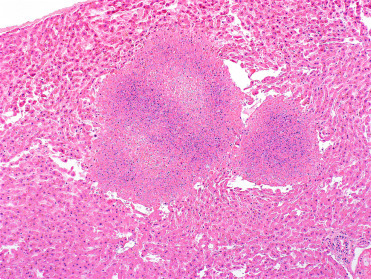
Tularemia in the liver of a free-ranging North American beaver.
Large, multifocal random areas of coagulative necrosis are present throughout the hepatic parenchyma. Note the relative lack of inflammation. Unlike Yersiniosis, bacterial colonies are not readily detected with routine hematoxylin and eosin staining.
(Photo Courtesy of D. Campbell, Canadian Wildlife Health Cooperative Ontario-Nunavut)