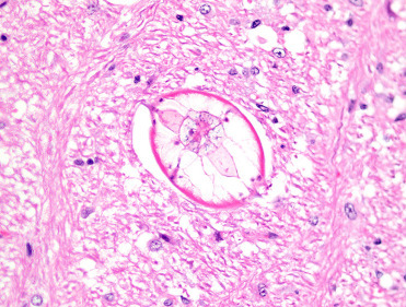
Figure 20.17.

Visceral larval migrans due to Baylisascaris procyonis in the brain of a free-ranging groundhog.
The larval nematode parasites contain prominent lateral alae and lateral cords, a thick cuticle, and coelomyarian musculature.
(Photo Courtesy of J. Landolfi, Of Mice and Microflora Considerations for Genetically Engineered Mice. P. M. Treuting, C. B. Clifford, R. S. Sellers, C. F. Brayton. First Published December 14, 2011)
