Figure 5.

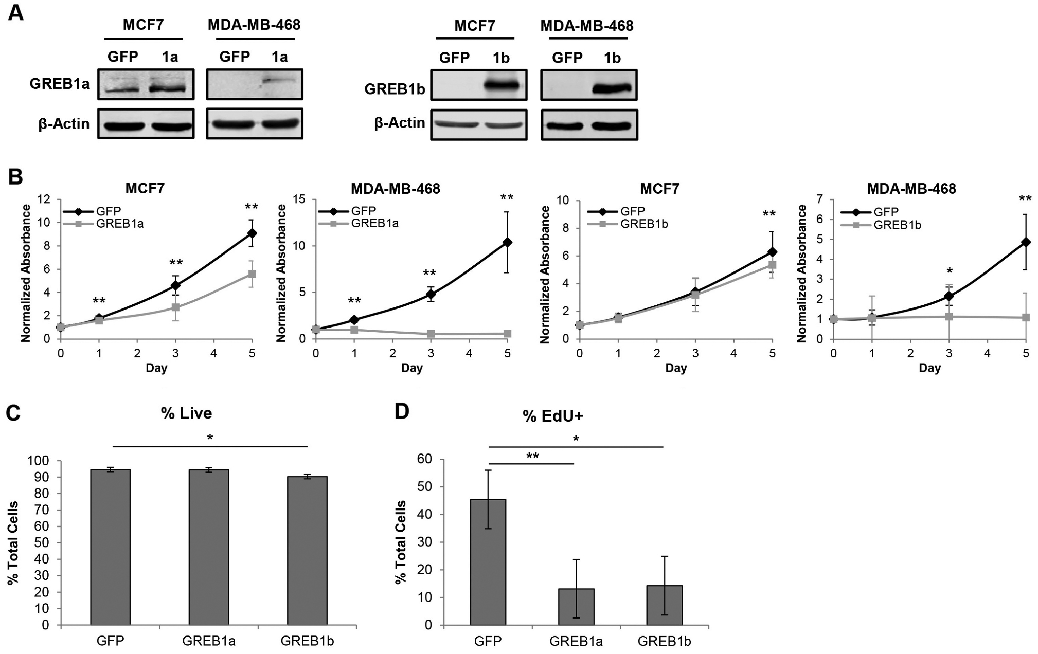
Elevated GREB1a and GREB1b expression reduces proliferation of breast cancer cells independent of ERα status. ER-positive (MCF7) and ER-negative (MDA-MB-468) cells were transduced with adenovirus expressing GFP, GREB1a, or GREB1b. (A) Immunoblot showing elevated expression of GREB1a and GREB1b in transduced cells compared to GFP-treated MCF7 cells. (B) Proliferation of MCF7 and MDA-MB-468 cells after transduction with GREB1a, GREB1b or GFP adenovirus was measured by MTT assay. Data are plotted as mean absorbance normalized to MTT Day 0 ± s.d.; n = 3 for each cell line. (C) MCF7 cells were transduced with GREB1a, GREB1b or GFP adenovirus. After 72 h, cells were stained with LIVE/DEAD Fixable Dead Cell Stain and analyzed by flow cytometry. Data are depicted as live cells as a mean percent total cells ± s.e.; n = 3. (D) MCF7 cells were transduced with GREB1a, GREB1b or GFP adenovirus. After 72 h, cells were treated with EdU and fixed after 6 h. Data are displayed as mean percent of EdU-positive cells ± s.e.; n = 3. *P ≤ 0.05, **P ≤ 0.01.
