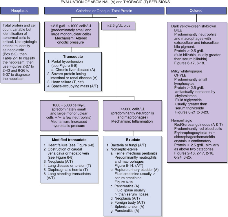
Figure 6-2.

Algorithm for effusion classification.
Abdominal and thoracic effusions are easily classified by color and content. Pathophysiology of the near colorless effusions depends on the etiology. Mild increases of cells and/or protein related to increased hydrostatic pressure (modified transudate) often result from chronic transudation of fluid or passage across a membrane. Exudation of significant numbers of neutrophils and/or macrophages from injured lymphatic and blood vessels (exudate) results from infectious or noninfectious etiologies. Colored effusions are distinctive forms of exudation.
(Modified from Meyer DJ, Harvey JW: Veterinary laboratory medicine—interpretation and diagnosis, ed 3, Elsevier, St Louis, 2004.)
