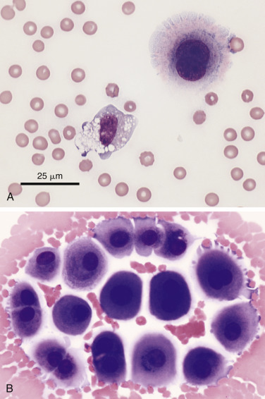
Figure 6-4.

Reactive mesothelial cell.
A, Exfoliated binucleate mesothelial cell (upper right) and mildly vacuolated and basophilic macrophage in an effusion. The mesothelial cell has a characteristic pink fringe along the cytoplasmic border. (Modified Wright; HP oil.) B, A loose group of variably reactive mesothelial cells at the feathered edge of a smear made from an effusion. These cells may contain one or more nuclei. Note the presence of the “fringe” (glycocalyx) on the mesothelial cells as well as prominent cytoplasmic blebbing of a few cells at the periphery. Several cells contain paranuclear dark granules, the significance of which is unknown. (Modified Wright; HP oil.)
