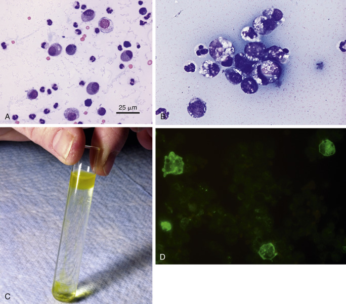
Figure 6-14.

Abdominal effusion. FIP. Cat.
A, This concentrated smear of the exudate contains moderately basophilic macrophages and nondegenerate neutrophils. The background contains basophilic, coarsely granular protein as well as basophilic protein crescents and strands of fibrin. (Modified Wright; HP oil.) B, This exudate mostly contains foamy, vacuolated macrophages with lower numbers of mildly degenerate neutrophils and intermediate to large lymphoid cells. Lymphocytes may be intermediate in size and appear reactive in some cases of FIP. Also note the granular precipitated protein throughout the background. (Methanolic Romanowsky; HP oil.) C, Rivalta test. Positive test results are indicated by a layer of gel on top of the acetic acid solution. Fluid is from a cat with PCR-confirmed FIP. The cat was moderately icteric. Note the yellow streaks of gel in the middle of the tube from partially floating material. D, FCoV immunofluorescence test. A specific cytologic test for FIP involves immunofluorescence of intracellular feline coronavirus (FCoV) within the effusion. Shown are three infected and intact macrophages (green). (FCoV immunofluorescence; HP oil.)
(C, Photo by Sam Royer, Purdue University. D, Courtesy of Jacqueline Norris, University of Sydney, Australia.)
