Figure 6-15.

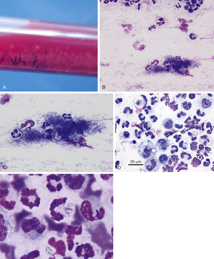
Septic exudate. Actinomycosis. Pleural. Same cat case A and E and same dog case B-D.
A, Gross fluid appearance showing presence of blood and numerous light yellow particles (i.e., sulfur granules). B, Direct smear from a pleural effusion demonstrates the cytologic appearance of a particle along with many lysed cells and nuclear streaks. These granules often are dragged to the feathered edge, as was the case with this smear. (Modified Wright; IP.) C, Higher magnification of the same particle in A. (Modified Wright; HP oil.) D, The fluid has marked suppurative inflammation with many degenerate neutrophils and several foamy macrophages. Short and long filamentous organisms and lysed cells are seen throughout the background. In areas of the smear lacking bacteria (not shown), the neutrophils were only mildly degenerate. (Modified Wright; HP oil.) E, Slender often beaded filamentous bacteria are present within two degenerate neutrophils. (Aqueous Romanowsky; HP oil.)
(A and E, Images courtesy of Janina Łukaszewska, Wrocław, Poland.)
