Figure 6-16.

Histoplasmosis. Dog.
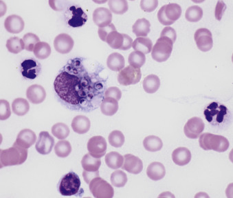
A macrophage containing numerous oval 2 to 3 μm yeast organisms of Histoplasma capsulatum is seen at left center. There are numerous red cells in the background of the smear. (Modified Wright; HP oil.)

Histoplasmosis. Dog.
A macrophage containing numerous oval 2 to 3 μm yeast organisms of Histoplasma capsulatum is seen at left center. There are numerous red cells in the background of the smear. (Modified Wright; HP oil.)