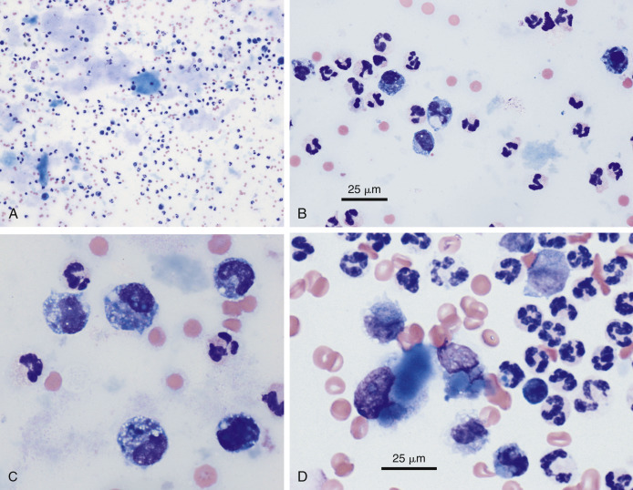
Figure 6-18.

Bile peritonitis. Dog. Same case A-C.
A, This is a concentrated smear of fluid from a dog with a ruptured gall bladder. Note the numerous neutrophils and lakes of blue-grey amorphous mucinous material that are present throughout the background. (Modified Wright; IP.) B, Note the suppurative inflammation with variably degenerate neutrophils as well as basophilic and foamy macrophages. Amorphous blue-gray material, likely mucin, is seen in the background. (Modified Wright; HP oil.) C, Note the suppurative inflammation and foamy macrophages that contain blue-grey to dark blue granular material. The background contains mucinous and finely granular protein. (Modified Wright; HP oil.) D, In abdominal fluid from another case, neutrophils appear mostly nondegenerate. Intracellular mucus material is dense, hyalinized, and medium blue. (Modified Wright; HP oil)
(D, Courtesy of Rose Raskin, Purdue University.)
