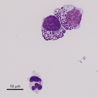
Figure 6-27.

Neoplastic effusion. Granular cell lymphoma. Pleural. Cat.
This fluid was light yellow, hazy with a protein of 4.2 g/dL and WBC of 5600/μL. In addition, 63% of nucleated cells were granulated; two are shown. Granules varied from fine to coarse (as shown) and were frequently eccentrically placed to one side of the cell. Nondegenerate neutrophils (one shown), small lymphocytes, and occasional phagocytes were also present. (Wright-Giemsa; HP oil.)
(Courtesy of Rose Raskin, University of Florida.)
