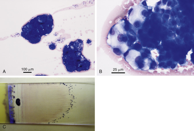
Figure 6-35.

Neoplastic effusion. Mesothelioma. Abdominal. Dog.
Same case A-C. A, One small cluster of reactive mesothelium is shown in comparison with two large neoplastic cell clusters. (Wright-Giemsa; LP.) B, Cell clusters are variable in size and cytoplasmic vacuolation. Morphology is indistinguishable from that of secretory adenocarcinoma. (Wright-Giemsa; HP oil.) C, Large cell clusters are grossly visible at the feathered edge of a direct smear of this abdominal fluid.
(Material courtesy of Sarah Hammond et al., Virginia Tech University; Case 6 of 2012 ASVCP case review session.)
