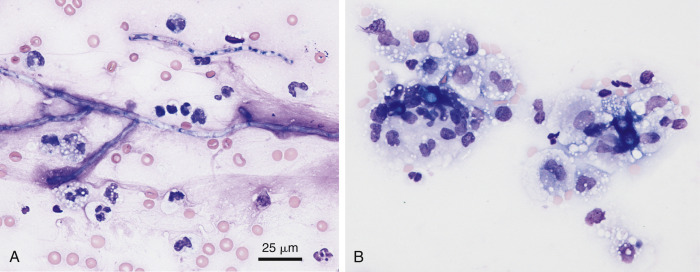
Figure 6-39.

Mixed cell fungal exudate. Pericardial. Dog.
A, Aspergillosis. Degenerative neutrophils and macrophages surround septate, branching hyphae of Aspergillus sp. as confirmed by fungal culture. (Modified Wright; HP oil.) B, Blastomycosis. Within the feathered edge of a direct smear, several dark blue fungal thick-walled yeast of Blastomyces dermatitides are surrounded by many vacuolated macrophages and small numbers of degenerate neutrophils. (Wright-Giemsa; IP.)
(A and B, Courtesy of Rose Raskin, Purdue University.)
