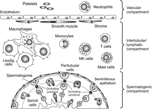
FIGURE 19.2.

Immunological compartmentalization of the testis.
The mammalian testis comprises three immunologically distinct compartments: the vascular compartment and intertubular (or interstitial) compartment are separated by a layer of nonfenestrated endothelium, while the intertubular and spermatogenic compartments are separated by a layer of peritubular myoid cells and by occluding junctions between adjacent Sertoli cells. These junctions constitute the blood–testis barrier, which further divides the seminiferous epithelium into a basal and an adluminal region (dotted line). The adluminal region contains the meiotic germ cells within a highly specialized microenvironment. Under normal conditions, monocytes, macrophages, T cells, NK cells and, in some species, mast cells and/or eosinophils have relatively free access to the intertubular compartment, but are entirely excluded from the adluminal region of the seminiferous epithelium. Neutrophils are confined to the vascular compartment except during specific immunological events.
