
FIGURE 23-19.
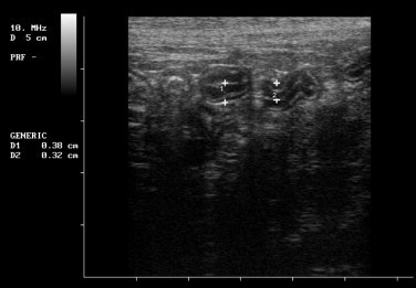
Ultrasonographic image of a cat with inflammatory bowel disease (IBD); the duodenal wall measurements at the two locations numbered D1 and D2 are 3.8 and 3.2 mm, respectively, compared with normal measurements of less than or equal to 2.8 mm. Note that most of the thickening is of the mucosal layer. The layers (from center) are lumen (white), mucosa (black), submucosa (white), muscularis (black), serosa (white).