
FIGURE 23-29.
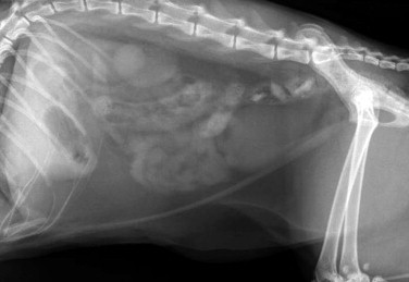
Radiographic appearance of a linear foreign body (lateral view). Note that most of the small intestine is localized in midventral region of the abdominal cavity instead of being dispersed uniformly throughout the peritoneal cavity.
Official websites use .gov
A
.gov website belongs to an official
government organization in the United States.
Secure .gov websites use HTTPS
A lock (
) or https:// means you've safely
connected to the .gov website. Share sensitive
information only on official, secure websites.
Radiographic appearance of a linear foreign body (lateral view). Note that most of the small intestine is localized in midventral region of the abdominal cavity instead of being dispersed uniformly throughout the peritoneal cavity.