
Figure 13-15.
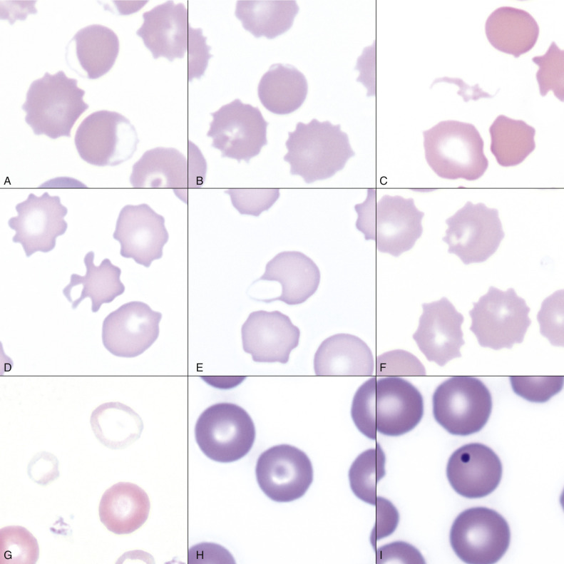
Common Erythrocyte Morphologic Abnormalities.
A, Blood from a dog that was administered a continuous rate infusion of propofol. The dog developed oxidant-induced hemolytic anemia with eccentrocytes. Wright-Giemsa stain. B, Blood from the same dog as in A, showing a pyknocyte. Note the spherocyte-like appearance of the pyknocyte, except for a small portion of the red blood cell membrane that is ruffled. Wright-Giemsa stain. C, A schistocyte in the blood of a dog with mechanical fragmentation hemolysis from disseminated intravascular coagulation. Wright-Giemsa stain. D, Blood from a dog with hemangiosarcoma, showing an acanthocyte. Wright-Giemsa stain. E, A keratocyte, exhibiting what appears to be a ruptured “vesicle” in blood from a dog. Wright-Giemsa stain. F, Blood from a dog with crenation artifact showing echinocytes. Wright-Giemsa stain. G, Blood from a dog with iron deficiency anemia. Note the patient's microcytic and hypochromic cell (left) and the normocytic hypochromic cell (top), as well as the normocytic normochromic erythrocyte (bottom right) from a recent blood transfusion. Wright-Giemsa stain. H, Blood from a dog. The center erythrocyte is a target cell, or codocyte. Wright-Giemsa stain. I, Blood from a dog shows a Howell-Jolly body, which is round, deeply basophilic remnant of the erythrocyte's nucleus. Wright-Giemsa stain.
(Courtesy Dr. K.M. Boes, College of Veterinary Medicine, Virginia Polytechnic Institute and State University.)